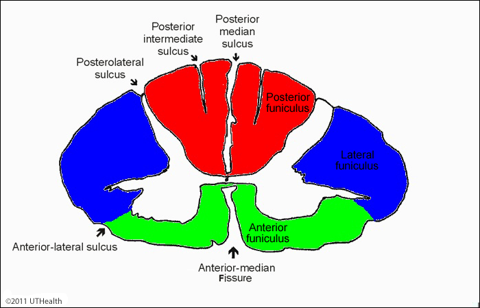
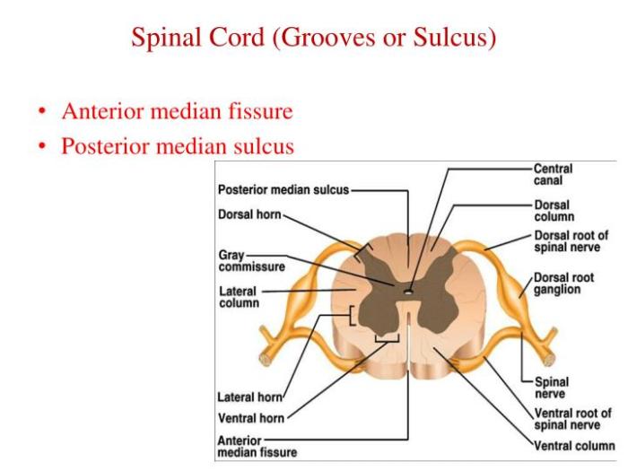
Posterior median sulcus spinal cord – The posterior median sulcus of the spinal cord is a pivotal anatomical structure that plays a critical role in understanding spinal cord function and pathology. Located at the dorsal midline of the spinal cord, this sulcus serves as a significant landmark for neurosurgeons and neurologists alike.
During embryological development, the posterior median sulcus forms through the intricate process of neural tube closure and differentiation of spinal cord tissues. This sulcus divides the posterior aspect of the spinal cord into two symmetrical halves, providing a clear demarcation between the left and right sides.
Posterior Median Sulcus

The posterior median sulcus is a deep, vertical groove located on the posterior surface of the spinal cord. It runs along the midline, dividing the posterior funiculus into two symmetrical halves.
The posterior median sulcus serves as an important anatomical landmark for identifying the posterior median septum, a thin sheet of connective tissue that separates the two halves of the posterior funiculus.
Anatomical Landmarks
- Posterior Median Septum:The posterior median septum is a thin sheet of connective tissue that separates the two halves of the posterior funiculus.
- Fasciculus Gracilis:The fasciculus gracilis is a white matter tract that carries sensory information from the lower body to the brain. It is located medial to the posterior median sulcus.
- Fasciculus Cuneatus:The fasciculus cuneatus is a white matter tract that carries sensory information from the upper body to the brain. It is located lateral to the posterior median sulcus.
Formation and Development of the Posterior Median Sulcus
The posterior median sulcus is a groove that runs along the dorsal midline of the spinal cord. It is formed during embryological development as a result of the closure of the neural tube and the differentiation of spinal cord tissues.
Neural Tube Closure
The neural tube is a structure that forms during early embryonic development. It is the precursor to the central nervous system, which includes the brain and spinal cord. The neural tube forms by a process called neurulation, which involves the folding and fusion of the ectoderm, the outermost layer of the embryo.
The neural tube closure begins at the rostral (head) end of the embryo and proceeds caudally (tailward). As the neural tube closes, it forms a groove along the dorsal midline of the embryo. This groove is called the neural groove.
Differentiation of Spinal Cord Tissues, Posterior median sulcus spinal cord
Once the neural tube is closed, the cells within the tube begin to differentiate into the various tissues of the spinal cord. The cells that line the neural tube form the ependymal layer, which is the innermost layer of the spinal cord.
The cells that surround the ependymal layer form the mantle layer, which contains the neurons and glial cells of the spinal cord. The cells that surround the mantle layer form the marginal layer, which contains the white matter of the spinal cord.
The posterior median sulcus is formed by the fusion of the neural folds along the dorsal midline of the embryo. The neural folds are the edges of the neural groove. As the neural folds fuse, they form a groove that runs along the dorsal midline of the spinal cord.
This groove is the posterior median sulcus.
Clinical Significance of the Posterior Median Sulcus: Posterior Median Sulcus Spinal Cord
The posterior median sulcus plays a crucial role in understanding spinal cord function. It serves as a boundary between the two dorsal funiculi, which carry sensory information from the body to the brain. Abnormalities in the posterior median sulcus can disrupt these sensory pathways, leading to neurological deficits.
Neurological Deficits Associated with Posterior Median Sulcus Abnormalities
Damage to the posterior median sulcus can cause a variety of neurological symptoms, including:
- Loss of fine touch and proprioception (position sense) in the upper extremities
- Impaired coordination and balance
- Spastic gait
- Charcot-Marie-Tooth disease, a genetic disorder characterized by progressive muscle weakness and atrophy in the hands and feet
- Syringomyelia, a condition in which a fluid-filled cavity forms within the spinal cord, causing progressive neurological deficits
Understanding the clinical significance of the posterior median sulcus is essential for diagnosing and managing these neurological conditions effectively.
Imaging Techniques for Visualizing the Posterior Median Sulcus

The posterior median sulcus can be visualized using various imaging techniques, including magnetic resonance imaging (MRI) and computed tomography (CT) scans.
On MRI, the posterior median sulcus appears as a linear hypointensity extending along the midline of the spinal cord on sagittal images. It is best visualized using T2-weighted sequences, where it appears as a dark line.
On CT scans, the posterior median sulcus is less conspicuous but may be seen as a thin, lucent line along the midline of the spinal cord on axial images.
Surgical Considerations Related to the Posterior Median Sulcus

The posterior median sulcus serves as a critical anatomical landmark during surgical interventions involving the spinal cord. Its precise identification facilitates safe and effective access to various regions of the spinal cord.
Surgical Procedures Utilizing the Posterior Median Sulcus
Knowledge of the posterior median sulcus is paramount in several surgical procedures, including:
Laminectomy
This procedure involves the removal of the lamina, the bony arch that forms the roof of the spinal canal. The posterior median sulcus serves as a guide for locating the midline and minimizing the risk of damaging the underlying spinal cord.
Spinal Cord Decompression
In cases of spinal cord compression due to herniated discs or tumors, the posterior median sulcus assists surgeons in identifying the affected area and performing a laminectomy to relieve the pressure on the spinal cord.
Myelomeningocele Repair
This surgery corrects a birth defect where the spinal cord protrudes through an opening in the vertebrae. The posterior median sulcus helps surgeons locate the defect and repair the spinal canal.
Spinal Cord Stimulation
In this procedure, electrodes are implanted near the posterior median sulcus to deliver electrical impulses to the spinal cord, providing pain relief for conditions such as chronic back pain.
Query Resolution
What is the clinical significance of the posterior median sulcus?
The posterior median sulcus serves as a crucial landmark for neurosurgeons during spinal cord surgeries. Its identification allows for precise localization of spinal cord structures and safe manipulation of the spinal cord during surgical procedures.
How is the posterior median sulcus visualized in medical imaging?
The posterior median sulcus can be visualized using various imaging techniques, including magnetic resonance imaging (MRI) and computed tomography (CT) scans. On MRI, it appears as a linear hypointense signal on sagittal and axial views, while on CT scans, it is seen as a thin, lucent line.